Enhanced Landmark Detection Model in Pelvic Fluoroscopy using 2D/3D Registration Loss
Overview
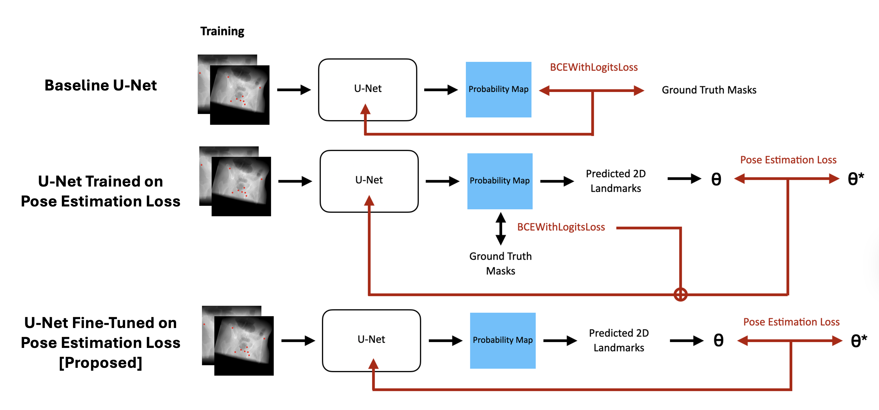
Anatomical landmark detection in pelvic fluoroscopy is critical for surgical planning in procedures like Total Hip Arthroplasty, where accurate localization guides component placement and biomechanical alignment. Existing U-Net-based methods assume a fixed Antero-Posterior view, which breaks down under realistic intraoperative conditions where patient or imaging unit orientation varies. We propose a training framework that integrates 2D/3D landmark-based registration into the U-Net training loop via a Pose Estimation Loss (PEL), penalizing geometric error between predicted 2D coordinates and ground truth 3D projections rather than relying solely on pixel-wise segmentation loss.
Key Contributions
- Proposed integrating 2D/3D registration as a Pose Estimation Loss into U-Net landmark detection training, directly penalizing geometric localization error.
- Demonstrated that sequential fine-tuning with PEL outperforms joint optimization, achieving 8.8% RMSE improvement on the external test set over the baseline.
- Showed that training with PEL alone diverges, and composite loss degrades performance (~2.4× error increase), establishing that pre-trained initialization is necessary for geometric loss to provide meaningful gradients.
Presentation Details
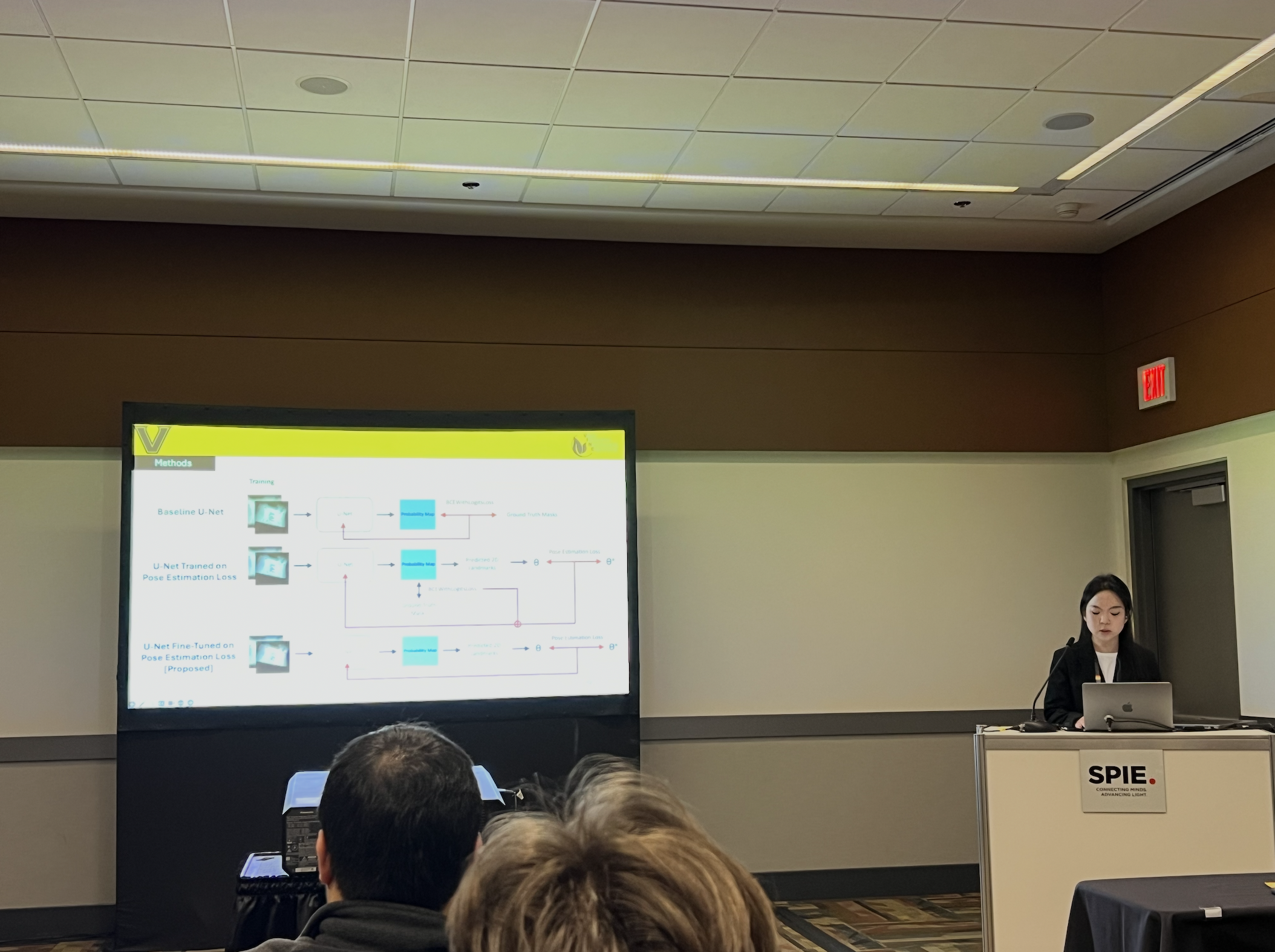
Presented as an oral talk at SPIE Medical Imaging 2026 in Vancouver, Canada. Work conducted at the VINE Lab (Vanderbilt University).